Search form

A.Z. Sint-Jan Brugge-Oostende AV campus Sint-Jan

Contact details
Traineeship proposition
Abstract
Testimony
Admin


Abstract Bachelor Project MLT 2020-2021:  Sensitivity determination of anaerobic germs using Micronaut-S microdilution

We evaluated the performance of the MICRONAUT-S microdilution broth assay, relative to the Antibiotic gradient test of Lyofilchem™, test strip by comparing the minimal inhibitory concentration (MIC) of different anaerobic bacteria against  different antibiotics. At present, according to European Society of Clinical Microbiology and Infectious diseases (EUCAST), there is no reference method for the antibiotic susceptibility assessment of anaerobes. Currently, the most commonly used method, the antibiotic gradient test strip, is laborious, time-consuming, burdensome for interpretation and reproducibility problems, which often hamper routine workflows. The microdilution method could be a great advantage in the routine, in terms of standardization and interpretation of the results. By using both tests simultaneously, the categorical and essential agreement between both methods were assessed. Furthermore, the hand on time, the practical and methodical advantages for both techniques for the lab technicians were determined. When the results of both methods are evaluated against the MIC values from reference isolates, it can be observed that the Micronaut-S showed better agreement than gradient test strip. For this kind of experiment, the repeatability, the reproducibility, the categorical and the essential agreement are above 90 percent (%) as required. When comparing between the two methods and the results versus the known MIC value, the CA is superior with the Micronaut-S than with the gradient strip test. The CA for the Micronaut-S at both ATCC and reference strains is 65.8% and for gradient strip it is 80.7%. Comparison relative to each other is 83.4%. Labor intensity and the hand on time is equal for both, also the purchase price is equivalent. The biggest difference would be in standardization, reading the results and interpretation, for which the Micronaut-S is considerably more straightforward.


Abstract 2020-2021 (advanced bachelor of bioinformatics): DEVELOPMENT OF A WEBTOOL FOR PROCESSING HIGH-THROUGPUT SEQUENCING DATA FOR SCREENING AND MONITORING PATIENTS AFTER A HAEMATOPOETIC STEM CELL TRANSPLANTATION
Patients suffering from certain haematological disorders may undergo a hematopoietic stem cell transplantation. At AZ Sint-Jan, site Bruges, pre- and post-transplant samples are taken and analyzed with a high-throughput sequencing based assay. The resulting data of screening (pre) and follow-up (post) samples are currently analyzed with the ADVYSER software (DEVYSER). This software calculates the host chimerism percentages for follow- up samples based on the sequence data. Drawbacks of the software are the lack of traceability, limited visualization capabilities hampering interpretation efficiency and reliability, and lack of networking possibility (the current access to the software is only possible via one computer in the lab). A new webtool was constructed to solve these disadvantages.
To construct the web-application and visualize the results, several computer languages are used. To construct the GUI a front-end open source toolkit, known as Bootstrap, was used. The HTML code, with styling via Bootstrap, was combined with PHP to upload and process data, and eventually display the results through a web interface. Uploaded data is stored partially in a MySQL database consisting out of two tables (screening and follow-up), and partially in a folder structure. Depending on the sample, data will be written to either the screening table or the follow-up table, and to specific files in the folder structure.
To facilitate interpretation, a preference was to show the different results in a more graphical way, as opposed to the ADVYSER software which provides a HTML text report only. To visualize the results on the webtool, several figures and tables are constructed for each sample type. This was performed via R. Upon file upload, a trigger will activate the R scripts and the figures are constructed.
Taking into account that mistakes may happen when uploading a sample, a function to edit or delete data was implemented. Via a webform the user can easily modify the information of a screening or follow-up sample, or delete an existing sample. To prevent major problems when performing this action, two checkups are implemented. A screening sample can only be modified if no follow-up sample is available. On the other hand, from a set of follow-up samples of a patient, only the most recent one can be modified. These different actions are all performed via HTML and PHP.
For easier navigation and adding more functionality to the web-pages, several Javascript functions were implemented. This was mostly done for showing different alerts and adding lookup tables for easier use of the webtool. According to these available functions, it can be concluded that the most important functions are already available. Yet the webtool is not final. Several aspects still need to be implemented. An example is the integration of an existing log-in system which will improve the traceability.
 
Abstract 2019-2020 (advanced bachelor of bioinformatics): Illumina run quality monitoring using R and a web GUI

For my internship I was asked to implement a web interface for monitoring quality parameters of Illumina runs. The data of these runs could already be visualized by the free application Illumina sequencing analysis viewer. This application has one major downside, it is not possible to compare values for the quality parameters between the different runs and samples. Being able to perform this on the web interface was the goal.

We worked in R to extract and read all the data for the runs. Using the packages XML and SavR these files could be handled in an R-script. SavR is used to parse and analyse Illumina SAV files, these files contain the quality parameters for one run. In the XML files, general info about the run can be found, such as run number and date. For some parameters extra steps were needed; calculating averages or discarding 0-values for example.

After processing all this data and calculating the needed values, these values are stored in a MySQL database thru the R-script. In the database we make a distinction between runparameters and sampleparameters. For the runs there are 23 different parameters in the database: RunID, Name, Total Reads, PF Reads, PF Percentage, Reads Identified, Q30, Density, PhasingR1, PhasingR2, PhasingR3, PhasingR4, PrephasingR1, PrephasingR2, PrephasingR3, PrephasingR4, Read1Length, Read2Length, Read3Length, Read4Length, Tiles, Date and Instrument. For comparing the samples only 4 parameters are used: RunID, Name, Fraction PF Reads and Reads.

Now that a database is created with all the data, we can call for these values, representing each run, to create plots and make it easy to compare different runs. For each parameter a separate script was created. In general, these scripts call for the info for each run in a loop for the corresponding parameter. After that a plot is made with this data and saved as an image. These images will be used in the web-interface.

For the actual web interface, we worked in html and php(notepad++). The bootstrap framework is used for designing the layout. It includes HTML and CSS based design templates and supports JavaScript plugins. In the php files a lot of interaction happen between the php file itself and the MySQL database.

The web interface contains 4 different start pages. The first one contains all info of the most recent run and comparison with the 20 previous runs. The second one is a list with all run numbers where the most recent one is on top off the page. The third one is the same but for samples. The last page is a list of instruments used between all the runs.

When clicking a run or sample on these pages, you get redirected to a details page for this particular run or sample. On this details page you can find a table with the values for all parameters. Underneath the table, you can find the comparison between the 20 previous samples/runs and the selected sample/run in the form of different graphs. Some graphs have cut-offs where a colour code can be applied. When clicking on an instrument, a list of run numbers is given. All these pages are linked together.

In the end we created a clear and simple web interface for comparing runs and samples. In addition an instrument page was created and the latest run has its own page. The script also runs automatically when a new folder with data is added to the parent folder.

Abstract Bachelor Project 1 MLT 2019-2020:  Validation of the BD BACTECTMFX for sterility testing of products from the bone and tissue bank and the hospital pharmacy

The current method of sterility tests at the pharmacy and tissue bank of the AZ Sint-Jan Hospital in Bruges shows a number of shortcomings such as the fact that it’s very labor-intensive. These sterility tests are used to verify the absence of viable and actively multiplying micro-organisms in preparations. This is done to prevent intravenous infections.

The plan is to replace the current manual method with an automated method using the BACTECTMFX Blood Culture System.

In this research, different pharmacy preparations (smoflipid/vitalipid, norepinephrine, growth 92 without trace elements and PCA epidural), stem cells and bone product are spiked with different types of micro-organisms. These suspensions are injected into a hemoculture bottle with a sterile syringe and needle. The hemoculture bottle is incubated in the BACTEC system until it’s flagged as positive which means that it can adequately detect growth. After this, micro-organisms are identified in the positive bottles. This identification is done with microscopy (Gram stain), culture and massaspectrometry (MALDI-tof).

In the first project, the bone and tissue bank project, the BACTEC method is compared to the standard plating method. This comparison led to the conclusion that the BACTEC method is faster than the plating method (2.56 vs 11.18 days). The BACTEC system is also the most sensitive and therefore the most accurate to identify C.albicans (53.85 vs 15.38%), as well as A.fumigatus (46.15 vs 38.46%) and Fusarium spp. (38.46 vs 0.00%). For the second project, with pharmacy preparations, the BACTEC system was validated in this study. The BACTEC method was found to be a sensitive method for different types of micro-organisms: S.aureus (100.00%), B.cereus (87.50%), P.aeruginosa (100.00%), C.albicans (87.50%) and A.fumigatus (75.00%). However it appeared to be less sensitive for anaerobes (C.perfringens 43.75% and B.fragilis 50.00%). This is not due to the BACTEC system but is caused by coming into contact with oxygen when making dilutions.

It can be concluded that the BACTEC system is a much easier and faster method with less hands on time, than the classic plating method when it’s used with suspensions that include bone product or stem cells. The method is very sensitive with pharmacy preparations as well. The current manual method can therefore be replaced by the automatic BACTEC method in the pharmacy and tissue bank of the AZ Sint-Jan Hospital in Bruges.

 
Abstract Bachelor Project 2 MLT 2019-2020:  Evaluation of automated tissue dissociation for flow cytometric analyses

The purpose of this research is to evaluate a new device (the MediMachine II) for automated tissue dissociation for flow cytometric analyses.

The current method for automated tissue dissociation is the GentleMacs Dissociator. Although already in use for several years, two main problems do occur. Sometimes an extra population is seen after an analysis, but there is no explanation for which cells this population indicates. The second problem is that sometimes malignant populations cannot be found with flow cytometric analysis. For these reasons, an alternative device will be evaluated (the MediMachine II). This device could be a solution for avoiding the two problems with the GentleMacs Dissociator.

To evaluate the device some parameters are examined. These are the correlation and difference in viability. These parameters will also be carried out with the manual method because it is known that with this method the least cells are destroyed. Also, the imprecision and the shelf life of a sample will be examined an as additional tests a price comparison will be made and the difference in ease of use will be examined. These tests will be done on lymph nodes and will be analyzed with the BD FacsCanto II flowcytometer.

The results of the tests show that the MediMachine II had higher correlation results for the analysis of CD19+. This can be important for analyzing B-cell chronic lymphoproliferative diseases. The difference in viability of the cells between the devices is notable. The stopping-gate of 5000 lymphocytes is more achieved with the MediMachine II than with the GentleMacs Dissociator. This may be due to the mechanical processing with the GentleMacs Dissociator, causing more destroyed cells. The imprecision is good with only a few values outside the limits. This is due to the heterogenicity of the tissue. Analyzing a lymph node is best within 24 hours. After that, the results can no longer be considered reliable. From the previous tests the MediMachine II is better but looking at the price and the ease of use, the GentleMacs Dissociator is better. The MediMachine II is more expensive and requires a manual and automatic processing step.

The results of the evaluation show that the viability of the cells is better with the MediMachine II and the manual method. This may be the reason that maligned populations with the GentleMacs Dissociator are more difficult to perceive. This parameter is an advantage for the MediMachine II. The switch to the MediMachine II will also depend on the other parameters because it’s more expensive and less easy to use.

 
Abstract Bachelor Project 3 MLT 2019-2020:  Evaluation of the use of IHC to detect MMR deficiency / micro satellite unstable tumors, in particular colorectal carcinomas

Lynch syndrome is an autosomal hereditary disorder that is the result of a mutation in one of the MMR-proteins (MLH1, PMS2, MSH2 and MSH6). Patients with Lynch syndrome have 25 to 70% chance to developing colon cancer. The aim of this research is to evaluate the correlation between micro satellite instability (MSI) and immunohistochemistry (IHC). There is also an evaluation of the reliability of a two- antibody panel.

MMR deficiency can be detected by immunohistochemical staining and / or MSI analysis. This study is about immunohistochemical staining. These stains are performed on the MSI panel of the Roche Ventana device. The indirect detection method is the method most frequently used on the Ventana.

There is a 98,37% correlation between MSI and IHC. There is 0,00% possibility of false positive results and a 1,63% possibility of false negative results. When there are ‘possible’ results, these tissue sections should be checked for any misinterpretation of the IHC because these tissue sections show abnormal expression. The Shia et al study confirmed that a two-antibody panel is as reliable as a four-antibody panel. Isolated loss of PMS2 and MSH6 gives a greater indication of Lynch syndrome than MLH1 and MSH2.

There is a good correlation between MSI and IHC. Both techniques will continue to be applied at AZ Sint-Jan in Bruges. From this study there can be concluded that the IHC is reliable if an MSI analysis confirms colorectal carcinomas. The four-antibody panel is still used. In the future, a two-antibody panel can be introduced in the lab.


Abstract Bachelor Project 1 MLT 2018-2019: The use of the Cobas® HPV test for detection of HR-HPV on FFPE in oropharyngeal tumors, a feasibility study
Human papillomavirus (HPV) not only causes cervical cancer, but also oropharyngeal cancer. The latter has been on the rise in recent years. Detection of HPV is now based on immunohistochemistry (IHC) and in situ hybridization (ISH), but these sometimes give unreliable results. That is why it is tested whether it can be detected by the Cobas 4800 HPV test from Roche which is a validated method for cervical and vaginal cytology.

Punching was performed from FFPE tissue from oropharyngeal tumors. The DNA was extracted using the MagCore, then diluted and placed on the Cobas. The results were compared with those obtained from the p16 IHC and HPV ISH, tested on the Benchmark Ultra from Ventana (Roche). Two methods for measuring DNA concentration were compared, showing that the Qubit is more reliable than the NanoDrop. By comparing different diluents, it was decided that the extract should be diluted in 1 ml of preservcyt or 1 ml of 50% ethanol. It was tested on two samples whether the extraction step with the MagCore could be omitted and could be extracted manually instead, but this must be further tested with more samples. The quality of the DNA was tested on the basis of a QuantiMIZE run. This test showed that the DNA from the oropharynx was damaged or too fragmented. Discrepant results were obtained for two of the ten HPV HR+ samples. Two of the ten HPV HR+ samples gave an invalid value. Some of the other 6 samples were positive for HPR16, but invalid for other HR HPV and HPV18. Invalid results were all obtained for the four oropharynx LR+ samples. The invalid values can be explained by the fragmentation of the DNA.

Invalid values and discrepant results were obtained for the oropharynx samples. This shows that the CE-IVD protocol on the Cobas 4800 for HPV testing is not a good method to detect HPV in oropharynx tumors.
 
 
Abstract Bachelor Project 2 MLT 2018-2019: Reducing the turnaround time of methicillin-resistant Staphylococcus aureus screening

Staphylococcus aureus is an important pathogen that can induce several types of bacterial infections. Over the past decades, there has been an alarming increase in methicillin-resistant Staphylococcus aureus (MRSA) strains.The aim of this study is to reduce the turnaround time (TAT) of the five-day MRSA screening in the AZ Sint-Jan Bruges hospital by using the Alere™ PBP2a Culture Colony Test, an immunochromatographic assay for rapid detection of methicillin resistance in bacterial colonies on culture media.

Over the course of eleven weeks, the test was performed on suspicious bacterial colonies from Oxoid™ Brilliance MRSA 2 media inoculated with patient samples. The results of the test were compared to the antimicrobial susceptibility test (AST) measured by the BD™ Phoenix™ M50. Additionally, the TAT after the introduction of this test was compared to the TAT of standard culture and AST screening.

Out of the 50 tested samples, 47 samples tested positive and three samples tested negative. One sample tested falsely positive compared to the AST. This resulted in a sensitivity of 100 % and a positive predictive value (PPV) of 97,87 %. Because of the selective components in the Brilliance MRSA 2 media resulting in only three negative test results, calculating the specificity and negative predictive value was not meaningful. The internal positive control of the test was positive for every test performed. Using the test resulted in an average TAT reduction of approximately one and a half days.

With a sensitivity of 100 % and a PPV of 97,87 %, the Alere™ PBP2a Culture Colony Test is suited for testing bacterial colonies from Oxoid™ Brilliance MRSA 2 media, resulting in an average TAT reduction of approximately one and a half days. After this study, use of the Alere™ PBP2a Culture Colony Test was implemented in MRSA screening in the AZ Sint-Jan Bruges hospital. In a future study, the impact of the TAT reduction on MRSA spread in the hospital could be investigated. Additionally, there could be experimented with reducing the incubation time of the tryptic soy broths before inoculating the Oxoid™ Brilliance MRSA 2 media.


Abstract Bachelor Project 3 MLT 2018-2019: Screening of organ-specific autoantibodies in liver diseases: a comparison of three liver/stomach/kidney substrates from the rat and the mouse with classical versus automated fluorescence microscopy

Indirect immunofluorescence assay (IFA) of patient serum on rodent tissue is a European standard for detection of autoimmune liver diseases which comprise autoimmune hepatitis (AIH), primary biliary cholangitis (PBC) and primary sclerosing cholangitis (PSC) as well as their overlapping syndromes. Early diagnosis using anti-smooth muscle (ASMA), anti-mitochondrial (AMA), anti-liver kidney microsome (LKM) antibodies as disease markers allows therapeutic intervention to prevent progression of these diseases. Stomach anti-parietal cell antibodies (APCA) in autoimmune gastritis can also be visualised on rodent tissue.

The aim of this study is to compare three rodent tissue kits: a mouse tissue kit from Menarini and a mouse and rat tissue kit from Werfen, to introduce this technique in the Sint-Jan Bruges hospital. For visualization of the autoantibodies, the classical fluoresence microscopy is compared with the automated fluorescence microscopy, except for the mouse tissue kit from Werfen which was analysed by classical microscopy.

During eight weeks, 50 samples were tested on the rodent tissue kits and analysed by different laboratory workers. The results were compared with those of the UZ Leuven hospital using the mouse Werfen substrate with classical fluorescence microscopy as reference method. Six samples with heterophilic antibodies were tested as well, since these samples are known to produce false positive results on the rodent substrate.

For ASMA, AMA, APCA and LKM antibodies 46 samples corresponded with the reference method using the mouse Werfen substrate and 41 samples with the mouse Menarini substrate, compared to 34 using the rat Werfen substrate. On mouse Menarini slides, three of the six heterophilic antibody samples scored negative, one was falsely classified as ASMA and in three of them no consensus was obtained. While on the Werfen mouse substrate, four samples were falsely classified as ASMA, one was negative and one without consensus. Five samples resulted in aspecific staining and one was without consensus on rat Werfen slides. The results from the mouse tissue from Menarini are about equal between manual and digital fluorescence microscopy. The results of the rat tissue from Werfen were much better with manual analysis than with digital fluorescence microscopy, reason being that there often was not enough rat tissue in the image of the screen for determination.

In conclusion, the results of ASMA, AMA, APCA and LKM antibodies on mouse tissue corresponded better with the mouse reference method than the results obtained on rat tissue. The mouse tissue from Menarini delivered the best result for heterophilic antibodies samples. Future investigations may focus on the comparison of the mouse substrate on the automatic microscopes of both companies.

 
Abstract advanced bachelor of bioinformatics 2018-2019BUILDING A PLATFORM FOR STORAGE, MANAGEMENT AND ANALYSIS OF VARIANTS DETECTED BY HIGH-THROUGHPUT SEQUENCING 
Introduction
In Belgium, until recently, there was no uniform, agreed-upon algorithm to determine the biological significance of a variant. In the Molecular Hematology lab at AZ Sint-Jan BruggeOostende, the significance of a variant is currently determined by a combination of 6 parameters: frequency in the COSMIC database, not frequently occurring in dbSNP/gnomAD/1000Genomes, SIFT classification, Polyphen classification, variant allele frequency (VAF) and whether or not the variant is located in a sequence encoding a conserved domain. Recently, a national work group has agreed upon an algorithm for the biological classification of variants. Based on these new rules, the biological class of a variant will be determined by the following parameters: frequency in the COSMIC database, classification as determined by SIFT, MutationTaster, JAX CKB, MDA, MCG, CIViCdb, ClinVar, OncoKB and Varsome, gnomAD frequency and evidence as available through PubMed. Each variant can be classified using this data according to a scoring algorithm. The goal of this is to make the process uniform across all hospitals in Belgium, so that the biological significance of a variant is the same in every hospital. The goal of the traineeship was to build a platform for storage, management and analysis of variants detected by high-throughput sequencing and the additional parameters required for biological classification. The three main pillars of this platform are 1. a MySQL database to store the variant data generated by the Illumina MiSeq, along with required parameters and metadata, 2. R scripts to parse the variant data files and annotate the variants in these files using information retrieved from online and offline databases, 3. a dynamic web tool written in HTML/CSS/JS/PHP which queries the MySQL database.
 
The MySQL database
Consequently, the blueprint of the database was determined by these new requirements. The main database consists of 7 tables: (i) a panel table which has the basic data of the different panels of genes; (ii) a run table which contains all the particulars from each time an experiment is run; (iii) a sample table which incorporates the necessary information of all the samples within the runs; (iv) a patient table which holds the patient details of the samples; (v) a variant table which stores all the variants that are detected by highthroughput sequencing; (vi) a variant-sample table which functions as a cross reference table between the sample and variant table; (vii) and finally a version table which shows when the annotation data of the linked databases was last updated. Additionally, there are 7 trace tables which log all the changes that are made to the 7 main tables, one for each, based on MySQL triggers. This is to ensure traceability of both automated and manual changes. Lastly, there are three tables for the ClinVar data: a ClinVar table with all the ClinVar information considered relevant, a PubMed table with the PubMed documentation associated with a variant and a ClinVar-PubMed table which functions as a cross-reference table between the former two.
 
R scripts for parsing and annotation
Using R scripts, the variant data files can be parsed and the important information can be extracted and inserted into the database using RMySQL. Other R scripts are then sourced to update the detected variants with additional annotation data from the genomic and other databases listed above. At this time, it is completed for COSMIC, dbSNP, SIFT, Polyphen, JAX CKB, ClinVar, gnomAD and PubMed. The majority of this information is retrieved through the Ensembl Variant Effect Predictor API. The NCBI Eutils API is used to retrieve ClinVar and PubMed data. gnomAD and JAX CKB data are downloaded and subsequently parsed. In the past, all the important variant data was manually curated in an Excel file. This old data also had to be inserted into the database. A separate R parsing script was created to extract all the useful information from the Excel table.
 
Graphical user interface
The web tool allows for the management and analysis of the variants stored in the database. It is mainly written in HTML, CSS and PHP, but also has some JavaScript for specific tasks. MySQLi prepared statements are used inside PHP to query the database. This results in a set of interconnected pages featuring the data from all the tables in the database which can be navigated through the navigation bar and the action buttons. The styling of the interface is created using Bootstrap.
 
Relevance
The benefit of this endeavor is that the information of the variant data files is now automatically parsed and stored in a database instead of it being manually inserted by a lab technician or a clinical biologist into an Excel table. This saves time and reduces mistakes due to manual curation, the storage system is more reliable and allows for tracing changes, and the web interface makes it easier to manage and analyze the variant data. 
 
Abstract bachelorproef 2017-2018: Co-testing of high risk human papillomavirus with the Cobas 4800 instrument

Cervical Cancer is one of the most common cancers in Belgium with 1414 new cases detected in 2011. The cancer is associated with the human papillomavirus (HPV) which is sexually transmitted. The traditional detection of cervical cancer is only based on the morphological image of the cells, but if the virus is latently present in the cells then is a morphological image unable to detect the virus. The advantages are an early detection of an HPV infection to start an early treatment, a better follow-up for the patient.

The aim of this thesis is to show the importance of co-testing. Co-testing is making a smear and colored with a Papanicoloau staining next to an HPV-testing on a sample with the Cobas 4800 from Roche independently of the result of the Papanicoloau staining.

If the smear is negative, the woman will be adviced to making every three years a new smear. Co-testing will also test for the HPV-DNA and when the sample contains HPV-DNA, the woman will be adviced to come back after one year for follow-up.

In this first part of the study is a global image about human papillomavirus, how works the traditional screening and what is co-testing.

In this second part of the study is tested the precision, reproducibility and a comparison of the positive NILM-samples between the Cobas 4800 and the validated MTA method.

The results in the study showed that 9,7% from the negative samples in the screening has a HPV-infection. There are 468 samples tested independent of the results of the screening (NILM, LSIL and HSIL). There is a 100% sensitivity and specificity with samples of the external quality control. Also, are there 93% of the HSIL samples positive for HPV tested with the Cobas 4800.

The Cobas 4800 is a good method for co-testing. When the comparison is tested between the Cobas and the MTA is the Cobas more sensitive for detect lower concentrations of HPV-DNA. The correctness is also good and is tested with external quality controls and with HSIL-samples.

 
Abstract bachelorproef 1 2016-2017: The evaluation of flow cytometric scoring systems for diagnosis of MDS

Myelodysplastic syndrome (MDS) are a group of clonal haematopoietic stem cell diseases characterized by peripheral cytopenia(s) and dysplastic morphology. The diagnosis relies on bone marrow (BM) cytomorphology and cytogenetics. However, recognition of dysplasia in BM smears can be challenging and cytogenetic abnormalities are not always found. On the other hand, the dysplastic features and cytogenetic abnormalities can also be found in conditions other than MDS and in healthy persons. So the diagnosis of MDS is not always straightforward. This thesis aims to evaluate the value of flow cytometry in the diagnosis of MDS.

First there was an evaluation of a flow cytometric score, the Ogata score. The four parameters used in this score are: myeloblasts(%), B-cell progenitors (%), myeloblast CD45 expression, and side scatter of granulocytes. These parameters can be determined with the Lymphoid Screening Tube (LST) that is analysed in every bone marrow sample of a patient without a history of a haematological disorder in AZ Sint-Jan. So, the Ogata score can be determined without an extra cost.

The second method was an extended Ogata score with CD56 expression on the myeloblasts and the monocytes (%). This parameters can also be determined with the LST.

For these methods a total of 37 MDS samples (22 low risk MDS and 15 high risk MDS) and 76 control patients with cytopenia were retrospectively analyzed.

For the third method a new MDS-panel was designed based on literature, in this panel the following parameters were used: CD45, CD15, CD56, CD34, CD11b, CD10, CD19 and CD7 each with a different function. For this prospective evaluation four MDS samples and four control samples were used.

The sensitivity of the Ogata score was 65% and the specificity 92%. For the extended Ogata score a sensitivity of 81% and specificity of 54% was found but their was a problem with an aspecific connection between CD56 and kappa on fluorochroom PE, result in false positive results. With the new MDS panel there wasn’t an added value with the Ogata score to distinguish MDS cases from control cases.

Flow cytometry can be used based on the standard Ogata score as a diagnostic tool for MDS. Because of the high specificity, the diagnosis can be suggested when a positive score is found. Moreover, the analysis can be performed without an extra cost.

Abstract bachelorproef 2 2016-2017Validation Optilite® CH50 Reagent on Optilite with reference method Wako® Autokit CH50 on P-Modular

Wegens confidentialiteit kan de samenvatting niet gepubliceerd worden.

Abstract bachelorproef 3 2016-2017Is the Idylla device of Biocartis effective for routine use in detecting EGFR / KRAS / BRAF mutations?

Wegens confidentialiteit kan de samenvatting niet gepubliceerd worden.

Abstract bachelorproef 1 2015-2016: Evaluation and comparison of four different analyzers for the quantification of HbA1c

To evaluate and compare four commercially available HbA1c analyzers (Menarini HA-8180, Bio-Rad D-100, Tosoh G8 and Sebia Capillarys 2 flex piercing) with respect to (1) correlation against the current laboratory HbA1c analyzer (Menarini HA-8160) and (2) imprecision against the HbA1c analytical goal of the coefficient of variation ≤ 2.9%.

Glycated hemoglobin (HbA1c) has become the preferred method for monitoring diabetes over the last decade. Various commercially available analyzers have been developed for measuring HbA1c in a routine laboratory. The analyzers should deliver fast and accurate results providing both the practitioner and the patient with the information for an appropriate diagnosis, treatment and follow-up of the patient.

The validation and comparison of the four analyzers are performed over a period of 2 weeks. During this period a series of standardized tests are executed and operator observations are recorded. The accuracy of each analyzer, including the laboratory analyzer, is assessed by using the calibrated SKML controls for both a low and a high HbA1c sample. The maximum allowed mean bias is 2 mmol/mol. The repeatability is performed by the consecutive measurement of ten low and ten high samples on each analyzer, whereas the reproducibility is executed by measuring ten identical both low and high samples twice a day during five consecutive days. For both criteria the calculated coefficient of variation CV must be maximum 2.9 %. The linearity is obtained by measuring test samples with linear increasing concentrations of 30 to 80 mmol/mol. A minimum R² of 0.95 is considered to pass this test. In order to verify the correlation of the new analyzers compared to the current laboratory analyzer (Menarini HA-8160) a series of 128 samples, including five hemoglobinopathy samples, is measured. Calculations and graphs of the linear regression with correlation coefficient r, the Intercept A, slope B at a confidence level of 95%, are performed using Passing Bablok regression and Bland Altman plot. Carry-over tests to check how efficiently the analyzer can manage eventual contamination by a previous high HbA1c sample on the next normal samples are executed. Lastly the functionality and the observations made while testing, in particular the features, the complexity, the required preparations, the quality of the reports, the software and the user interface are evaluated.

The coefficient of variation is acceptable for all four analyzers, although the CVs of the repeatability and the reproducibility for both the Menarini HA-8180 and the Tosoh G8 analyzers are significantly lower than for the two other instruments. In respect to linearity only Tosoh G8 (R² = 0.947) is slightly below the target. The Capillarys of Sebia shows the best linearity (R² = 0.9988). Passing-Bablok regression executed on the four analyzers compared with the current Menarini HA-8160 shows only for Sebia an intercept A outside the confidence interval of 95%. Carry-over does not appear on either analyzer.

The analyzer with the best user interface, the least complexity and generating the fastest and the best reports is the Bio-Rad D-100 analyzer.  

The Menarini HA-8180 is the best choice considering all criteria with acceptable precision, good linearity and acceptable agreement with the central laboratory results. The more only this instrument provides the feature of obtaining the HbA2 concentration simultaneously with the HbA1c measurement. Also in regard to simplicity of operation, fast and reliable reporting, the Menarini HA-8180 scores high.

 
Abstract bachelorproef 2 2015-2016Evaluatie van drie HPLC’s en één capillaire elektroforese voor analyse van hemoglobinopathieën 

In the AZ Sint-Jan Hospital Campus Brugge an HPLC system (Menarini HA-8160) is used for analysis of different types of hemoglobin: HbA1c, HbA0, HbA2, HbF and hemoglobin variants. With this system different clinical manifestations can be detected such as monitoring of patients with diabetes by the quantification of HbA1c and detection of hemoglobinopathies (thalassemias and hemoglobin variants) by quantification of HbA2, HbF and any hemoglobin variants. After thirteen years of duty, this system needs to be replaced, therefore an evaluation of three new HPLC systems and one capillary electrophoresis is performed on demo systems to decide what system would replace the currently used one. In this thesis the evaluation of the four systems for analysis of hemoglobinopathies is displayed. The three HPLC systems are Menarini HA8180T, TOSOH G8 and Biorad D-100. The capillary electrophoresis system is Sebia Capillarys. These where tested on quality control, between run, intra run, correlation, linearity, carry-over and advantages and pitfalls for using them. During these tests 64 blood samples where used, these contained samples with α-and β-thalassemia’s, and hemoglobin variants such as HbA/S, HbS/S, HbA/E, HbA/D-punjab, HbA/C, HbA/Lepore, HbA/J-baltimore and HbA/Muravera. The results showed that every system had its pitfalls and its advantages, although there were no major deviations with clinical significance. This means that in the end every system has successfully passed the evaluation.

Abstract bachelorproef 3 2015-2016Comparing study of two methods for the determination of high-risk HPV

Worldwide there were more than 500.000 new diagnoses of cervical cancer in 2012. With a mortality of about 50%, 250.000 women die of this disease annually. Over 99% of these cancers are caused by high risk human papilloma viruses. The quicker the treatment, the better the prognosis. Therefore, the early detection of the virus is very important.
The main purpose of this study is to compare the Cepheid Xpert HPV-assay and the Hologic Cervista HPV-HR test. Both methods test for high risk HPV on cervical PAP-smears. The comparison include hands-on time, runtime, verification and workload.
The Cepheid company has provided 30 HPV high risk screening tests. From our recent files 30 PAP-samples were selected upon which Cervista HPV high risk tests were already performed. The 30 samples include five ASC-US, five NILM, five LSIL and five HSIL-samples. The reproducibility will be tested by testing a proven HPV-positive sample three times on the Cervista MTA and six times on the GeneXpert. The different modules of the GeneXpert will be tested separately.
The HPV-typing of both methods have about the same result. The MTA has a higher sensitivity, sometimes generating false positive results. Because the GeneXpert is single-sample based, the results are immediately available, but generate a higher cost per test. The choice between the two methods depend on the amount of samples that need to be tested. The more samples, the better the MTA is for a technique. In case of only a few samples a day the GeneXpert is preferred, but with a higher cost.

 
Samenvatting eindwerk 1 2014-2015: THE SWEAT TEST AND ITS ALTERNATIVES IN THE DIAGNOSIS OF CYSTIC FIBROSIS
Aim – This research project presents a two-fold aim. Firstly, two methods for sweat collection will be compared, practically and analytically: the current Gibson-Cooke method (GCM) and the Macroduct Sweat Collection System (MSCS). The main objective of this method comparison is to determine whether or not these two methods are interchangeable. Secondly, the performance of the Chlorochek Chloridometer will be determined. If the machine is considered performant, it could be stated as a good replacement for the older machine that is currently in use (Chloride Analyzer 925) in the AZ Sint-Jan Hospital in Bruges.
Background – Sweat chloride is increased in patients with cystic fibrosis (higher than 60mmol/L). The sweat test is the golden standard in the diagnosis of cystic fibrosis. The current sweat collection method, the Gibson-Cooke procedure, show multiple cons (risk of evaporation, poor accuracy, time-consuming). The Macroduct Sweat Collection System claims to rule out all of these disadvantages. The Chloride Analyzer 925 is currently used to determine the chloride concentration in sweat samples. This device has been used for decades and the company doesn’t offer any support when having technical issues. Therefore, a reliable replacement device is needed.
Experimental Design – By using different levels of Sweat Controls different performance criteria of the Chlorochek Chloridometer will be determined (trueness, bias, accuracy, storage conditions, linearity and LOQ). Secondly, twenty healthy volunteers will undergo the sweat test using both techniques. Sweat will be analyzed on both devices. The results of the method comparison will be processed using Passing and Bablok regression and Bland-Altman plots.
Results – Practically, more problems could be observed using the MSCS in comparison with the GCM. The MSCS also had a much higher failure rate (35%). The Chlorochek Chloridometer had an acceptable total error of 6,41% and 4,13% when using Sweat Controls level 2 and 3. When using level 1, the total error was too high (19,92%), but this is clinically irrelevant in the lower range. Storage in the refrigerator is recommended. The linear range goes from 10 to 100mmol/L. The LOQ is at 10mmol/L. Regarding Passing and Bablok regression, GCM/925 is analytically the most interchangeable with the GCM/Chlorochek method. The Bland-Altman plot reveals a mean difference of 0,7 mmol/L between this two methods which can be neglected according to the RCPA criteria.
Conclusion – The Gibson-Cooke method will be further used in combination with the new, performant device, the Chlorochek Chloridometer.
 
Samenvatting eindwerk 2 2014-2015: Optimization viability tests with flow cytometry
The purpose of this research comes from two known problems in the flowcytometry lab within the field of hematology. The first problem arises when samples arrive on Friday evening and they aren’t analyzed until Monday. To maintain a stable sample through the 72h span, we investigated what the benefit would be of adding a fixative, medium or preservative.
A new transport fixative has been tested with different kinds of samples and should be able to bridge a maximum of seven-ten days, guarantying sample integrity. There is also a preservative that guarantees stabilization of samples up to seven days.
For the stability tests six bronchoalveolar lavage (BAL) fluids, two peripheral bloods, two pleura fluids and two bone marrows are analyzed with and without fixatives. One gland is analyzed with and without a preservative and a medium. The samples will all be analyzed respectively at 0h, 24h, 48h and 72h through a screening tube with the BD Fluorescence Activated Cell Sorter (FACS) Canto II flow cytometer.
The results of the stability tests show that the results without adding stabilizers give equal or better results until at least 72h. The use of fixative with BAL-fluids shows various results, but the correlation between both methods is high. The use of preservative gives good results for the pleural fluids, peripheral blood samples and bone marrows. The correlation between both methods is high and the maximum deviation of 10% is never exceeded, except with 7-AAD. The viability is expected to lower through the time that passes. The results of the gland seem to be diverse. With the preservative, the correlation is very low and the maximum 10% is mostly exceeded. The use of RPMI shows a much higher correlation and almost never exceeds the 10% deviation. Overall, the results for the method with or without stabilizers are both accepted for clinical diagnostics. With this in mind, the stabilizers do not give an added value at different time points.
The second problem lies within the difficulty in differentiating live from dead cells by using the viability marker 7-AAD. Viability assays are performed on flow cytometric analysis of body fluids, glands and cryopreserved stem cells.
The current viability marker 7-AAD provides an unpredictable discriminative power, which makes the gating problematic. Within this problem there is a need for an alternative viability marker. In an abstract (Moshaver, B., 2013), the viability markers 7-AAD and DRAQ7 were compared, resulting with a higher discriminative power for DRAQ7 and a higher non-specific binding for 7-AAD.
Because of these findings a new method was created to analyze samples with the new viability dye DRAQ7. For the viability tests four glands, four fluids and five stem cell samples are analyzed with 7-AAD and DRAQ7. The samples, except the stem cells at 0h (hours), will all be analyzed respectively at 0h, 24h, 48h and 72h through a screening tube with the BD FACSCanto II flow cytometer.
The results of the viability tests for the glands and pleural fluids show that the use of DRAQ7 has a better discriminative power in comparison to 7-AAD. The other markers do not seem to be influenced within the dot-plots by the compensation needed for the use of DRAQ7. 7-AAD will be replaced with DRAQ7 for the tests of glands and fluids. The results of the thawed stem cells show that the use of DRAQ7 in the FACSCanto software has a greater discriminative power in comparison to 7-AAD. The problem within this research is that the absolute counting of the white blood cells and stem cells do not match the reference values. With this in mind, a switch from 7AAD to DRAQ7 does not seem feasible for the analysis of cryopreserved stem cells.
 
Samenvatting eindwerk 1 2013-2014: Optimalisation and standardization of flow cytometric diagnostics with EuroFlow-panels and Infinicyt Software
Flowcytometric immunophenotyping is a simple and fast technique used for identification, classification and follow-up of different hematologic malignancies, such as acute myeloid leukemia (AML), acute lymphoid leukemia (ALL), myelodysplasia (MDS) and different Non-Hodgkin Lymphomas (NHL). In recent years, a lot of efforts have been made to standardize flowcytometric analysis. Euroflow is an organization that has developed standardized flowcytometric protocols, antibody panels and software (Infinicyt). The purpose of this work is to compare three Euroflow flowcytometric panels to the current panels used in the hematology lab of AZ Sint-Jan: the Lymphoid Screening Tube (LST), the Acute Leukemia Orientation Tube (ALOT) and the AML/MDS panel. Also, the Infinicyt software will be evaluated.
For the LST tube three panels were evaluated: the current panel (containing CD34) and two Euroflow-approved panels, both of which contain the same markers (including CD5), but for one tube the antibodies have to be pipetted separately, while for the other tube (ready-to-use Cytognos kit) only one pipetting step is necessary. The Cytognos kit also contains TCRgd. There were only minor analytical differences between the current panel and the two Euroflow panels. Practically, it was decided that the Cytognos kit was too expensive. Also, CD34 is important in screening for determination of blast percentage and CD5 for screening for monoclonal B-lymphocytosis. It was decided to use the Euroflow LST tube with CD5 for screening in peripheral blood and the current LST tube with CD34 for screening in bone marrow..
For the ALOT tube three panels were evaluated: the current panel and two Euroflow-approved panels (one with separate pipetting of the antibodies and one ready-to-use Cytognos kit). The three panels contain the same markers, but the two Euroflow-panels contain Euroflow-approved clones of the antibodies. In the Euroflow-approved panels, expression for cyCD79a and CD19 was stronger, resulting in a better discrimination between the negative and positive population. Again, the Cytognos kit is too expensive, so it was decided to switch to the other Euroflow ALOT tube for routine analysis.
For the AML/MDS panel, the Euroflow panel was compared to the current AML/MDS panel. There were only minor analytical differences between the two panels. However, the Euroflow panel contains 9 new valuable markers. It was decided to switch to the Euroflow panel for routine analysis, although it is more expensive than the current panel. However, the price can be reduced by analysing the most specific markers (ie for megakaryoblastic or erythroid leukemia) only when morphologically indicated.
The Infinicyt software offers the possibility to build databases and draw maturation pathways of normal samples. Pathological samples can then be compared to these pathways. Due to some technical difficulties and the experience needed for working with this software, we were not able to build this database. Further evaluation of this software will be necessary in the future.
 
Samenvatting eindwerk 2 2013-2014: Validation of the RAS mutation analysis
In the laboratory of pathological anatomy at the AZ Sint-Jan in Bruges, Three kits from the firm Qiagen are used for the quantitative detection of mutations in RAS-genes. The Therascreen KRAS Pyro kit and Therascreen NRAS Pyro kit are used for the analysis of codon 12, 13 and 61 in the KRAS- and NRAS-gene. The RAS Extension kit is used for the analysis of codon  58, 59, 117, 118, 146 and 147 in both genes.
The laboratory is already accredited for the mutation analysis of codon 12, 13, and 61 in the KRAS-gene. For the comprehensive analysis the lab isn’t accredited.
In order to obtain accreditation, the method should be validated. To prove how reliable and sensitive the test is, some validation parameters are required to be determined, namely:
  • Sensitivity
  • Reproducibility
  • Repeatability
  • Cut- off value
  • Linearity
 
The validation process is divided in two runs. The first run will function as a ‘in-between’ run. The results from the samples are known before the start of this run. In this run the reproducibility can be determined. The second run can be used as a ‘intra’-run. Here the repeatability can be determined. Because the second run consists a serial dilution, the cut-off value can be defined.      
All samples are treated as the same way as in the laboratory pathological anatomy.
Each sample undergoes the following steps:
  • Dilution of the DNA extract to a DNA concentration of 2ng/µl.
  • ‘Polymerase chain reaction’ step: specific DNA sequences are amplified to generate enough DNA, necessary for analysis.
  • Pyrosequencing with the PyroMark Q24: with this method, the sequence of nucleotides in DNA can be defined. Mutations can be detected in the RAS-genes.
All the validation parameters are valued in a positive way. The reproducibility and repeatability are both 100%. The cut-off value satisfies the conditions provided by the firm Qiagen. The linearity is sufficiently reliable.
 
Samenvatting 1 eindwerk 2012-2013: Validatie van von Willebrand Factor antigen en activiteit op ACL TOP 700
Het doel van deze bachelorproef is het evalueren van alternatieve assays om von Willebrand Factor (VWF) antigen en activiteit te bepalen. De VWF is een belangrijk onderdeel van zowel de primaire als de secundaire hemostase. Deze factor helpt bij het herstellen van een beschadigde bloedvatwand door samen met de bloedplaatjes een trombocytenplug te vormen en functioneert als een transporteiwit voor stollingsfactor VIII. Het belang van het bepalen van von Willebrand Factor antigen en activiteit situeert zich in het opsporen van de ziekte van von Willebrand. Deze (erfelijke) bloedingsziekte wordt verdeeld in drie grote categorieën: een partiële kwantitatieve deficiëntie van VWF (type 1), een kwalitatieve deficiëntie (type 2) en een totale deficiëntie van VWF (type 3).
Binnen de bachelorproef wordt gezocht naar een alternatief ter vervanging van de huidige meetmethodes in het AZ Sint-Jan, campus Brugge. De huidige methode voor activiteitsbepaling, aggregometrie, is arbeidsintensief en weinig reproduceerbaar. Voor de reagentia van de huidige methode voor antigenbepaling (VIDAS, Biomérieux) heerst momenteel een productiestop, waardoor een alternatief noodzakelijk is. In dit eindwerk werd de bepaling van VWF antigen en activiteit via latex-immunoturbidimetrie geëvalueerd op de stollingsautomaat ACL TOP 700 (Instrumention Laboratory). Voor VWF activiteit werd zowel een kit met ristocetine, als een kit zonder ristocetine uitgetest. De evaluatie omvat volgende parameters: precisie (herhaalbaarheid en reproduceerbaarheid), juistheid (externe kwaliteitscontroles en correlatie met de gouden standaard), correlatie met de huidige methode, lineariteit en controle van de referentiewaarden. De referentiewaarden voor personen met bloedgroep O voor VWF antigen en activiteit zijn respectievelijk 42,0 – 140,8% en 48,2 – 201,9%. Voor personen met bloedgroep A + B + AB ligt het interval iets hoger, namelijk 66,1 – 176,3% VWF antigen en 60,8 – 239,8% VWF activiteit.
oostende
Samenvatting 2 eindwerk 2012-2013: Validatie van de EGFR mutatie analyse bij niet kleincellige longcarcinomen
EGFR is een receptor aanwezig op het celmembraan die vele aspecten van het cellulair metabolisme reguleert waaronder de celproliferatie, de celdifferentiatie en de celoverleving. Door een mutatie in het EGFR gen zal de receptor continu signaaltransductiemechanismen activeren die leiden tot een verhoogde proliferatie van de cel. Deze mutatie is bij 15% van de niet kleincellige longcarcinoma’s aanwezig waarbij het opsporen van deze mutatie belangrijk is in het kader van de behandeling. Wanneer een mutatie in het EGFR gen aanwezig is kan een behandeling gestart worden met specifieke TKI’s. Deze TKI’s zullen de receptor blokkeren door binding op de actieve site van de receptor.
Het doel van deze bachelorproef is om een EGFR mutatie analyse methode te valideren. Men gebruikt hiervoor de therascreen EGFR Pyro kit van de firma Qiagen. Voor deze kit op basis van pyrosequencing moet de accuraatheid tegenover een referentie, de reproduceerbaarheid en de cut-off waarde berekend en beoordeeld worden.
De analysemethode bestaat uit verschillende stappen. Eerst wordt in formol gefixeerd en in paraffine ingebed weefsel uitgesneden op basis van een haematoxyline-eosine coupe. DNA extractie wordt vervolgens uitgevoerd op het uitgesneden weefsel. Na een verdunning van de verkregen hoeveelheid DNA wordt het geviseerde stuk DNA geamplificeerd met de PCR methode. Daarna wordt het geamplificeerde DNA gescheiden van onzuiverheden en wordt er enkelstrengig DNA gevormd waarop de pyrosequence analyse gebeurt. Het resultaat is een pyrogram die de sequenties en eventuele mutaties weergeeft.
De accuraatheid van deze methode wordt berekend door het opnieuw uitvoeren van mutatieanalyses op tien stalen die eerder getest zijn in een referentielabo. Bij deze stalen wordt steeds hetzelfde resultaat bekomen als in het referentielabo waardoor de accuraatheid van de test op 100% kan gelegd worden.
Voor de berekening van de precisie word één staal elf maal gemeten. Hierbij ziet men dat steeds hetzelfde resultaat bekomen wordt bij de mutatieanalyses, waardoor men een precisie uitkomt van 100%.
De cut-off wordt bepaald door het maken van een verdunningsreeks op twee stalen. Hierbij heeft de ene een mutatie in exon 19 en de andere een mutatie in exon 21. Bij het bekijken van de pyrogrammen kan de cut-off waarde voor exon 19 gelegd worden op een mutatiegraad van 4,1% en voor exon 21 op 6,5 % mutatiegraad, wat voldoet aan de vooropgestelde cut-off waarden gegeven door de firma namelijk 4.2% voor exon 19 en 8.5% voor exon 21.
Hieruit kunnen we besluiten dat de kit voldoende accuraat, precies en gevoelig is om de aanwezigheid van een mutatie aan te geven. De therascreen EGFR pyrosequencing kit van Qiagen is dus een voldoende gevoelige en betrouwbare manier om EGFR mutaties op te sporen.
 
Samenvatting eindwerk 1 2011-2012: Validatie van de BRAF mutatie analyse op melanoma
Een naevus, een goedaardige proliferatie van melanocyten, is een benigne pigmentcelletsel. Onder bepaalde omstandigheden kan deze naevus ontaarden in een maligne melanoom. De alarmtekenen voor deze ontaarding worden weergegeven in de ABCD-regel. Een melanoom kan echter ook de novo ontstaan. In bepaalde gevallen is er een mutatie in melanomen. Volgens de literatuur blijkt dat er in 66% van de melanomen de BRAF mutatie aanwezig is. In deze mutanten is er tot 92% de BRAF V600E mutatie.
Het doel van deze bachelorproef is om de BRAF mutatie analyse methode te valideren. Hiervoor moet de cut-off waarde, de accuraatheid tegenover een referentie en de reproduceerbaarheid berekend en beoordeeld worden. De analysemethode bestaat uit verschillende stappen. Eerst wordt in formol gefixeerd en in paraffine ingebed weefsel uitgesneden op basis van een H&E coupe. Hiermee wordt de DNA extractie uitgevoerd. Na een verdunning van de DNA concentratie wordt het geviseerde stuk DNA geamplificeerd met de PCR methode. Daarna wordt het DNA gescheiden van onzuiverheden en wordt er enkelstrengig DNA gevormd waarop de pyrosequence analyse gebeurt. Het resultaat is een rapport met pieken dat de sequentie weergeeft.
 
Samenvatting eindwerk 2 2011-2012: Validatie van automatische celtelling in verschillende lichaamsvochten op de Beckman Coulter UniCel® DxH 800
Het manueel tellen van rode - en witte bloedcellen in lichaamsvochten is zeer arbeidsintensief en zeer variabel. Deze twee factoren kunnen verholpen worden door lichaamsvochten automatisch te laten tellen door een toestel. In deze thesis wordt de Beckman Coulter UniCel® DxH 800 besproken.
De Beckman Coulter UniCel® DxH 800 werkt volgens het impedantieprincipe. Hierbij wordt de verandering van elektrische weerstand gemeten wanneer er een gekernde cel of rode bloedcel voorbij de meetopening komt. De verandering van elektrische weerstand komt overeen met de grootte van de cel, uitgedrukt in femtoliter. Met behulp van voorgelegde grenswaarden voor gekernde cellen en rode bloedcellen kan hierdoor een onderscheid gemaakt worden tussen gekernde cellen en rode bloedcellen.
Om een nieuw toestel in de routine te plaatsen, moet er allereerst een validatie gebeuren op het toestel. Een validatie is namelijk nodig om te zien of het toestel wel correct meet. Als “gouden standaard” wordt het manueel tellen van lichaamsvochten met behulp van een microscoop gebruikt. De resultaten van de Beckman Coulter UniCel® DxH 800 worden vervolgens vergeleken met deze van de manuele telling. Tijdens de validatie wordt de precisie, nauwkeurigheid, lineariteit, houdbaarheid, ‘carry over’ en de controle van de referentiewaarden bepaald. Hierbij worden er performantievoorwaarden vooropgesteld waar de resultaten aan moeten voldoen.
Bij de validatie werden er verscheidene lichaamsvochten gebruikt. Het meest geanalyseerde lichaamsvocht is lumbaalvocht. Deze is zeer belangrijk doordat het een kritische grenswaarde heeft van vijf witte bloedcellen per µl. Naast lumbaalvochten werden pleuravochten en ascitesvochten het meest geanalyseerd, vervolgens broncho-alveolaire lavagevochten , continue ambulante peritoneale dialysevochten  en pericardvochten.
De automatische celtelling, uitgevoerd door de Beckman Coulter UniCel® DxH 800, toonde een slechte correlatie met de manuele telling bij lumbaalvochten. Dit komt doordat er onder het meetbereik van het toestel wordt gewerkt. Aangezien de kritische grens bij vijf witte bloedcellen per µl ligt, werd aan de hand van de resultaten van deze studie beslist de lumbaalvochten in de toekomst niet automatisch te tellen op de Beckman Coulter UniCel® DxH 800. Andere lichaamsvochten vertoonden een redelijke tot goede correlatie tussen de automatische celtelling en de manuele telling, uitgezonderd in het geval wanneer het aantal cellen per µl groter is dan het meetbereik van het toestel. Deze zullen ook automatisch op de Beckman Coulter UniCel® DxH 800 worden geteld. Indien het aantal getelde cellen in het lichaamsvocht lager is dan deze van het meetbereik van het toestel, dan wordt het lichaamsvocht nog manueel geteld. Tot slot om ‘carry over’ te vermijden, wordt er steeds een blanco gemeten tot deze een ‘total nucleated cell’ meetwaarde weergeeft van minder dan twintig per µl en er minder dan 1000 rode bloedcellen per µl aanwezig zijn.
 
Samenvatting eindwerk 1 2010-2011: BRAF mutatie analyse op punctiecytologie in diagnostiek van papillair schildkliercarcinoom
Het BRAF eiwit is een kinase dat betrokken is in een belangrijke pathway voor de transductie van mitogene signalen van de celmembraan naar de kern. Door een mutatie in het BRAF gen ontstaat een continu expressie van het BRAF eiwit wat kankervorming tot gevolg kan hebben. De BRAF mutatie wordt dan ook regelmatig teruggevonden bij verschillende kankers, vnl. bij coloncarcinoma, melanomen en papillair schildkliercarcinoma.
In deze studie wordt het zoeken van de BRAF-mutatie beperkt tot papillaire schildkliercarcinoma. In het archief worden papillaire schildkliercarcinomen gezocht waarvan paraffineblokjes met ingebed weefsel voorhanden zijn. Naast deze paraffineblokjes is ook cytologisch materiaal beschikbaar dat bekomen werd door een fijnenaaldaspiratie op de schildklier. Zowel bij de paraffineblokjes als bij het cytologisch materiaal wordt met behulp van de therascreen BRAF pyro kit (Qiagen) gezocht naar de mutaties.
Uit voorgaand wetenschappelijk onderzoek werd aangetoond dat de mutatie op twee verschillende gebieden kan voorkomen nl. op codon 600 en op de codons 464-469. Op de verschillende stalen worden respectievelijk een DNA extractie, PCR en een pyrosequencing analyse uitgevoerd. Deze technieken laten toe om de mutaties in codon 600 en codons 464-469 op te sporen. Op 45% (5/11) van de biopsiestalen is de mutatie van codon 600 aangetoond. In de codons 464-469 is geen mutatie gevonden. De literatuur bevestigt dat de mutatie in codon 600 frequenter voorkomt, namelijk voor 80%.
Bij het cytologische materiaal wordt geen mutatie teruggevonden.
Geen van de onderzochte stalen was cytologisch maligne of verdacht van maligniteit. Dit werd namelijk opgespoord door een DLP uit te voeren in het verleden. In het cytologisch onderzoek werden geen maligne cellen teruggevonden. Om te kunnen weten of het opsporen van de BRAF mutatie in cyologisch materiaal mogelijk is, wordt in de toekomst een methode gevolgd die met zekerheid maligne cellen geeft in een cytologisch staal. Daarom wordt momenteel  op verse resectiestukken die in het labo binnenkomen gepuncteerd zodat met zekerheid maligne cellen aanwezig zijn in het cytologisch materiaal.
Het vinden van de BRAF-mutatie is vooral belangrijk voor de therapie. Door een constante activatie van het BRAF-eiwit is het toedienen van een behandeling veel nuttiger bij gemuteerde patiënten dan bij niet-gemuteerde. Door dit eindwerk wordt bewezen dat het opsporen van de mutaties zeker een belangrijke rol gaat spelen in de toekomst en via verder onderzoek zal dit ook kunnen gebeuren op cytologisch materiaal zodat de last voor de patiënt beperkt blijft.
 
Samenvatting eindwerk 2 2010-2011: Gevoeligheidsbepaling met de VITEK 2 Compact (BioMérieux), Phoenix (Becton Dickinson) en E-testen (BioMérieux, Lucron)
In deze bachelorproef worden verschillende methoden van microbiële gevoeligheidsbepaling met elkaar vergeleken.
Het gaat om een vergelijking tussen twee geautomatiseerde methoden (de VITEK 2 Compact en Phoenix) en een manuele methode: de Etest® van BioMérieux en de MIC Test Strip® van Lucron. De werkwijze en principes van deze methodes worden besproken. Deze vergelijking wordt uitgevoerd op vijf bacteriën: Staphylococcus aureus, MRSA, Escherichia coli, Haemophilus influenzae, Acinetobacter baumannii en Streptococcus pneumoniae.
In het geval van H. Influenzae worden de E-testen vergeleken met een ander toestel: de Mini API. Dit komt door het feit dat H. Influenzae in de routine niet getest wordt op de VITEK 2 Compact. Voor H. Influenzae worden er ook twee verschillende Mueller Hinton Fastidious bodems getest en vergeleken. Het gaat hier om de Mueller Hinton Agar with 5% horse blood + 20 mg/l β-NAD® van BioMérieux en de Mueller Hinton Fastidious Agar® van Lucron
Het eindwerk geeft een overzicht van het klinisch belang, biochemische eigenschappen,  antibiogram en resistentiemechanismen bij deze kiemen. De volgende antibiotica worden gebruikt en vergeleken: vancomycine, cefoxitine, amoxicilline-clavulaanzuur, piperacilline-tazobactam,           temocilline, cefuroxime, ampicilline en penicilline.
De werking van deze antibiotica wordt samen met resistentiemechanismen besproken in een apart hoofdstuk.
De testen worden wanneer mogelijk uitgevoerd volgens de EUCAST richtlijnen. De EUCAST richtlijnen zijn de meest recente breekpunten die gepubliceerd zijn. De E-testen zijn in batch ingezet om ervoor te zorgen dat de resultaten vergelijkbaar zijn. Deze resultaten worden in tabelvorm naast elkaar gezet en geïnterpreteerd.
Uit de resultaten van deze testen kan men afleiden dat er onderling tussen de verschillende E-testen weinig grote verschillen in interpretatie te zien zijn. Het grootste verschil tussen de twee merken is het feit dat de Etest® heteroresistente kolonies opgemerkt heeft, terwijl de MIC Test Strip® deze niet opmerkt.
De toestellen vertonen echter verschillende discrepanties tegenover elkaar. Verschillende, reeds gekende problemen met amoxicilline-clavulaanzuur en piperacilline-tazobactam zijn te zien in de resultaten en worden verder besproken. Vancomycine geeft op de VITEK 2 Compact bij een MIC-waarde van 2 een onbetrouwbaar resultaat dat geconfirmeerd moet worden.
Naast deze gekende problemen komen de VITEK 2 Compact en de Phoenix goed overeen op vlak van interpretatie.
De problemen met de Mini-API zijn duidelijk en E-testen kunnen een vervangmiddel zijn voor dit toestel.
De resultaten van de vergelijking tussen de  Mueller Hinton Agar with 5% horse blood + 20 mg/l β-NAD® en de Mueller Hinton Fastidious Agar® komen grotendeels overeen. Praktisch gezien geeft dit soort platen soms moeilijk af te lezen E-testen. De bodem is minder doorzichtig, en H. influenzae is over het algemeen een moeilijke groeier. Indien de kiem goed groeit zijn de platen wel gemakkelijk af te lezen. De Mueller Hinton Fastidious Agar® had echter wel één groot nadeel: de platen waren vochtig. Dit kan een negatief effect hebben op E-testen.
Praktisch gezien zijn de E-testen gemakkelijk in te zetten, maar wel arbeidsintensief. De MIC Test Strip® heeft enkele kleine praktische voordelen tegenover de Etest®.


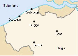
Address

Ruddershove 10
8000 Brugge
Belgium
Ruddershove 10
8000 Brugge
Belgium

Contacts

Martine Lansens
martine.lansens@azstjan.be
Dr. Ivo Van Den Berghe
Dr. Jacques Van Huysse
jacques.vanhuysse@azsintjan.be
Stefanie Ketelers
Dr. Jan Emmerechts
Dr. Michel Langlois
M. Vercammen
Stefanie Vermeire
Matthijs Vynck
matthijs.vynck@azsintjan.be
Katelijne Floré
katelijne.flore@azsintjan.be
Eric Nulens
eric.nulens@azsintjan.be
Zoekopdracht
Klassiek
Via Map